Part 6. How Does Dyskinesia Develop?
— In a disease defined by too little dopamine, why does the body sometimes move too much?
Parkinson’s disease is commonly described as a disease of reduced movement.
The hands slow, steps shorten, the body stiffens, and initiating movement becomes difficult. So when first learning about Parkinson’s disease, it is natural to think:
“It’s a disease where too little dopamine makes movement difficult.”
But after many years of disease and long-term levodopa treatment, the opposite can appear.
The body cannot stay still. The neck and shoulders sway, the trunk twists, and the arms and legs move involuntarily as if dancing.
This is levodopa-induced dyskinesia (LID).
It seems paradoxical.
The disease began with too little dopamine, and after replacing that missing dopamine, movement can become excessive.
For a long time, I thought of this simply as “the medication hitting too strongly.” But studying neuroanatomy and pharmacology again changed that view.
Dyskinesia is not simply caused by too much dopamine.
What matters more is how dopamine is delivered to the brain over time.
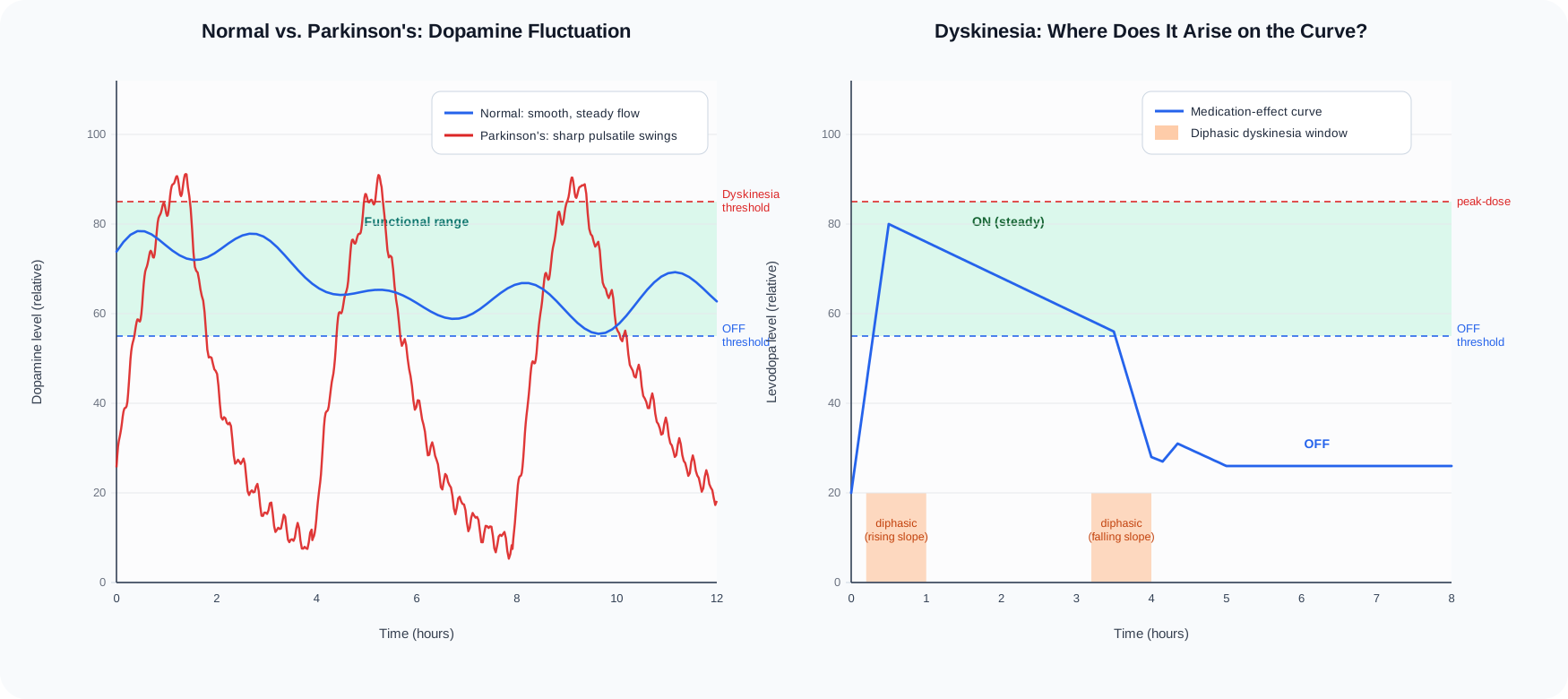
Figure. Normal buffering smooths dopamine delivery. As buffering capacity is lost, plasma levodopa fluctuations are transmitted more directly to the striatum, and dyskinesia may appear at the peak or during rising and falling phases.
1. In the normal brain, dopamine flows relatively smoothly
Dopamine neurons in the substantia nigra continuously send dopamine to the striatum.
The concentration does not simply surge and collapse. Instead, a relatively steady baseline is maintained, with smaller changes added as needed.
A simple analogy is a faucet that keeps water flowing steadily.
Whenever we move, the system makes fine adjustments such as:
“Move now.”
“Maintain this movement.”
“Stop now.”
The basal ganglia are therefore not simply an ON/OFF switch for movement.
They are closer to
a system that continuously fine-tunes the size and speed of movement.
2. In Parkinson’s disease, the neurons that store and buffer dopamine are lost
In Parkinson’s disease, dopamine neurons in the substantia nigra pars compacta gradually disappear.
Early in the disease, surviving neurons can still compensate to some degree.
Levodopa entering the brain can be converted to dopamine, stored, and released in a more regulated way.
That is one reason medication effects can feel relatively smooth early on.
But as more dopamine neurons are lost, the situation changes.
The ability to store and buffer dopamine declines.
As a result, changes in plasma levodopa concentration are transmitted more directly to the striatum.
A useful analogy is a reservoir.
Earlier in the disease, excess input could be stored and released more gradually.
As the reservoir shrinks, medication produces a sharper rise, and when the effect falls, dopamine signaling falls more sharply as well.
3. The problem is the pulse, not just the amount
After an oral levodopa dose, blood levels rise and then fall.
Taken several times a day, this produces repeated oscillations.
When enough dopamine terminals remain, those oscillations can be buffered.
When many have been lost, striatal dopamine receptors repeatedly experience:
strong stimulation → weak stimulation → strong stimulation → weak stimulation
This is often called pulsatile dopaminergic stimulation.
I think this is one of the key ideas for understanding dyskinesia.
Rather than being simply
a state of too much dopamine,
it is better understood as
an abnormal motor circuit created as the brain adapts over time to excessively fluctuating dopamine signaling.
4. The direct pathway can begin to open too far
The basal ganglia contain the direct pathway, which facilitates movement, and the indirect pathway, which suppresses movement.
Dopamine activates the direct pathway through D1 receptors and reduces the suppressive effect of the indirect pathway through D2 receptors.
Overall, dopamine makes movement easier.
In the OFF state, dopamine is deficient.
The direct pathway weakens and inhibitory influences become stronger.
So
movement becomes difficult.
When levodopa acts strongly, however, the direct pathway can become activated very rapidly.
After long exposure to pulsatile stimulation, striatal neurons can become abnormally sensitive.
The movement-promoting signal can then become stronger than necessary.
The body is, in effect, receiving too much of the command:
“Move.”
5. The problem does not end with the D1 receptor
Dyskinesia cannot be explained by saying only that D1 receptors are stimulated.
Many intracellular signaling systems sit downstream of dopamine receptors.
One commonly discussed cascade is:
D1 receptor → cAMP → PKA → DARPP-32
With repeated strong dopaminergic stimulation, these signaling systems can become hypersensitive, and even gene expression and synaptic properties can change.
The brain is not simply reacting to the drug in that moment.
Over time, repeated stimulation can cause
the circuit itself to learn and change.
This can be understood as a change in neuroplasticity.
Plasticity is useful in normal learning.
In dyskinesia, however, the system may have learned in the wrong direction.
6. Glutamate is pulled into the instability as well
The basal ganglia do not run on dopamine alone.
The major excitatory signal from cortex to striatum is glutamate.
When dopamine signaling repeatedly fluctuates, glutamatergic signaling changes as well.
Changes in the function and localization of NMDA and AMPA receptors can increase the excitability of striatal neurons.
So dyskinesia can be viewed as
not only a dopamine disorder, but a circuit disorder produced together by dopamine and glutamate.
This also helps explain why amantadine can reduce dyskinesia.
Amantadine reduces excessive NMDA receptor-mediated glutamatergic signaling.
7. What happens at the output of the basal ganglia during dyskinesia?
The GPi, or globus pallidus internus, is one of the major output structures of the basal ganglia and normally exerts tonic inhibition on the thalamus.
When the direct pathway is activated, GPi activity falls.
The inhibition on the thalamus is then released.
This is called disinhibition.
Once freed from excessive inhibition, the thalamus can drive the motor cortex more strongly.
A moderate amount of disinhibition allows normal movement.
Too much can produce excessive motor cortical output.
The result can be
unwanted movement, excessive trunk movement, and involuntary limb movement,
that is, dyskinesia.
OFF and dyskinesia may look like completely different states, but they can be seen as opposite ends of the same circuit.
In OFF, the gate is too closed.
In dyskinesia, the gate is too open.
8. Why can dyskinesia appear when the medication is working best?
The most common pattern is peak-dose dyskinesia.
It appears when levodopa effect is near its highest level.
The experience can be strange.
The body moves well.
Walking is easier, the hands work better, and rigidity loosens.
But at the same time, the body sways or writhes.
It can be described as
ON, but an excessively high ON.
My own experience of dyskinesia has also been difficult to reduce to only two states, ON and OFF.
A day can look more like:
OFF → rising phase → good ON → dyskinesia → good ON again → falling phase → OFF
The height and direction of the medication-effect curve both matter.
If Parkinson’s disease is recorded only as
ON / OFF,
much of this information disappears.
9. Dyskinesia can also appear while medication is rising or falling
Not all dyskinesia is peak-dose dyskinesia.
There is also diphasic dyskinesia, which appears during intermediate phases as levodopa effect rises or falls.
In this pattern, abnormal movement may be most evident
while moving into ON, or while falling back toward OFF,
rather than at the peak.
This means that in some patients,
reducing medication can paradoxically worsen dyskinesia,
while a small additional levodopa dose can sometimes reduce it.
That is one reason dyskinesia management is not simple.
Visible excessive movement should not automatically lead to the conclusion:
“Too much medication.”
The first question is
where on the medication-effect curve did the dyskinesia occur?
10. Dyskinesia and dystonia overlap, but they are not the same
Another common problem in Parkinson’s disease is dystonia.
The toes may curl inward, the foot may twist, or a specific muscle group may contract in a sustained way.
Dyskinesia is often more flowing and irregular.
Dystonia more often produces sustained contraction and fixed abnormal posture.
But in real patients, the two are not always cleanly separated.
During rising or falling transition phases,
dyskinesia and dystonia can overlap.
The body does not always follow textbook categories neatly.
11. In the end, the goal is not to give as much dopamine as possible
The goal of Parkinson’s medication treatment is not simple.
If levodopa is continually increased to eliminate OFF, dyskinesia may worsen.
If medication is reduced too aggressively to eliminate dyskinesia, severe OFF may appear.
So the goal is neither
to give as much dopamine as possible
nor
to reduce dopamine as much as possible.
What matters most is:
keeping dopaminergic stimulation as stable as possible within the range the brain can tolerate.
This is one of the most important principles I have learned from repeatedly recording my own medication response.
Sometimes lowering the trough matters more than raising the peak.
Good treatment may not be about making the highest point higher.
It may be about
flattening the medication-effect curve.
Dyskinesia, understood through my own body
For a long time I thought of dyskinesia as simply
“a state of too much medication.”
Now I see it differently.
As Parkinson’s disease progresses, the brain loses some of its ability to store and buffer dopamine.
Each levodopa dose can then produce a sharper rise and fall in dopaminergic stimulation.
With repetition, striatal circuits become progressively more sensitive.
Eventually,
a circuit that should produce normal movement can begin to produce more movement than necessary.
Dyskinesia is therefore not a side effect that appears overnight.
It is a change in neural circuitry created by long disease progression together with repeated dopaminergic stimulation.
To understand dyskinesia, one pill is not enough.
We have to look at
the entire dopamine curve over time.
That is why, in recording my own Parkinson’s disease, I kept returning to the rise and fall of medication effect, the peaks and the troughs.
Part 6 in one line
Dyskinesia is not caused simply by too much dopamine. It develops as repeated fluctuations in dopaminergic signaling reshape the basal ganglia motor circuit into a hypersensitive state.