Part 2. The basal ganglia — the system that selects movement
For a while, I understood Parkinson’s disease this way:
Dopamine is lacking, so the body slows down.
Not wrong.
But watching my body over a long time, another question arose.
The legs still have strength — why won’t the first step come?
Once walking starts, I can walk — why is setting off so hard?
On flat ground my stride shrinks — why, in front of stairs, does my step suddenly grow?
These questions eventually made me look past dopamine itself to the circuit dopamine acts on.
At its center sit the basal ganglia.
Chapter 9. Where are the basal ganglia?
The basal ganglia are a collection of nuclei deep beneath the cerebral cortex.
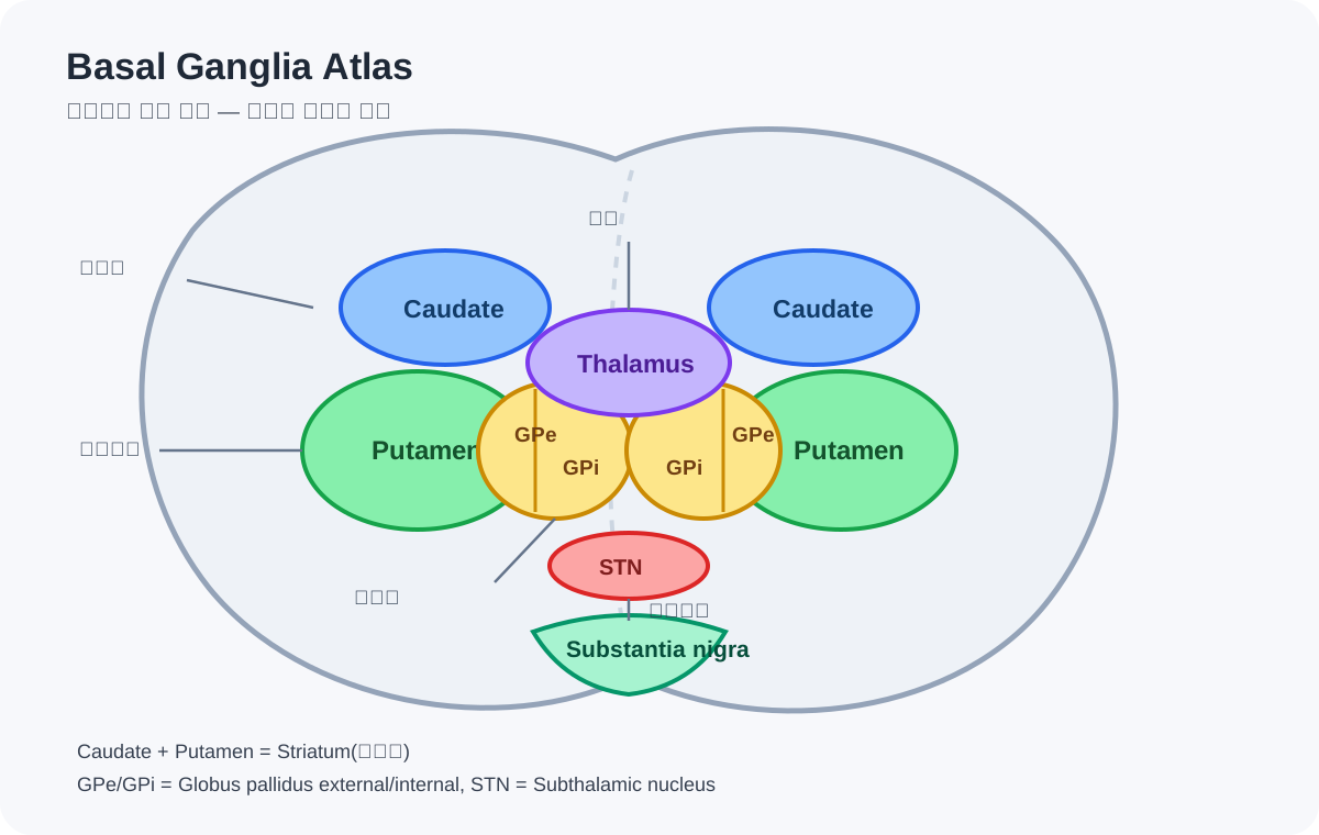
The core of the motor circuit is formed by the striatum, the globus pallidus, the subthalamic nucleus (STN), and the substantia nigra.
The striatum divides into the caudate and the putamen.
For movement, the putamen matters most.
The figure below is a simplified educational atlas of how these structures sit in relation to one another.
At first the names look daunting.
But once you grasp the direction of the circuit, it becomes surprisingly simple.
Information enters from the cerebral cortex → the striatum receives it → selection and suppression are regulated inside the basal ganglia → the output leaves through GPi/SNr to the thalamus → and returns to the motor cortex.
The basal ganglia converse with the cortex continuously, like a closed loop.
Chapter 10. The striatum — the doorway where movement enters
Motor-related information generated in the cerebral cortex enters the basal ganglia.
Its main doorway is the striatum.
In the motor circuit, the putamen is especially important.
What arrives here is not only the movement being attempted, but sensation, habitual movements, and the context of preceding actions.
I found it easiest to picture the striatum as the reception desk where movement candidates arrive.
Start walking.
Turn the body.
Rise from the chair.
Reach out a hand.
Many possible actions exist at once, yet we select one and execute it.
The basal ganglia take part in exactly that selection.
So when the basal ganglia go wrong, even without outright paralysis, the process of drawing a movement out can become difficult.
Chapter 11. The basal ganglia’s output is, at baseline, closer to a brake
Here you meet a fact that seems odd at first.
The basal ganglia’s main output nuclei, GPi and SNr, act to continuously inhibit the thalamus.
Put simply, they keep a baseline brake on, so that no movement pops out at random.
To make a movement, that brake must be released appropriately for the intended action.
Meanwhile, unnecessary movements must stay suppressed.
So the basal ganglia cannot be captured by a single accelerator-or-brake metaphor.
More precisely, they are
a selection system that lets the needed movement through while suppressing its competitors.
Seen this way, Parkinsonian bradykinesia also looks different.
It is not that the muscles cannot generate force — it is that the needed motor program is not being released enough.
Chapter 12. The direct and indirect pathways
Study the basal ganglia and you will inevitably meet the direct and indirect pathways.
At first there are too many arrows to memorize.
But the core question is single:
“In the end, how much should the thalamus be inhibited?”
The direct pathway works to reduce GPi/SNr inhibition for the needed movement.
The thalamus becomes relatively free, and activating the motor cortex becomes easier.
The indirect pathway, conversely, contributes to suppressing competing or unnecessary movements.
The STN holds an important position here, raising the output of GPi/SNr.
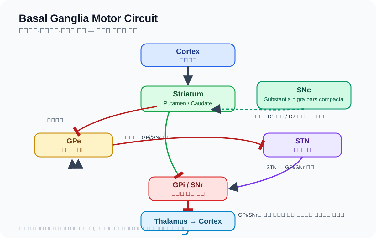
Rather than memorizing this figure, it connects better to real movement to understand it as a system that opens the gate for the needed movement and closes the gates on its competitors.
Chapter 13. What does the dopamine of the substantia nigra do?
The structure most often mentioned in Parkinson’s disease is the substantia nigra.
In particular, the dopamine neurons of the substantia nigra pars compacta (SNc) are lost.
These neurons send dopamine to the striatum.
Dopamine is not simply a substance that switches movement “on.”
Acting differently on the D1-type neurons of the direct pathway and the D2-type neurons of the indirect pathway, it tilts the circuit’s balance toward a state where the needed movement is more easily selected and executed.
So when dopamine runs short, the muscles do not simply weaken.
The basal ganglia’s output can tilt toward over-suppressing movement.
As a result, the signal returning to the motor cortex struggles to reach sufficient size and timing.
In my body, it felt like this:
I can move — but movement does not come out easily.
Chapter 14. Why does even the size of movements shrink?
Calling Parkinsonian bradykinesia merely “slowness” misses something important.
Steps shorten.
Handwriting shrinks.
Arm swing diminishes.
Repeated movements grow progressively smaller.
This connects to the fact that the basal ganglia take part not only in selecting movements but in setting their size and vigor.
That is why a conscious external instruction —
“One big step.”
“Swing your arms wide.”
— can enlarge the movement for a while.
A conscious target stands in for the size that used to be set automatically.
This was a crucial hint for me.
Moving small is not only a matter of weak muscles.
The circuit that automatically sets movement size from within is being shaken.
Chapter 15. Why is the first step the hardest?
I have the will to walk.
I know where I am going.
My legs have strength.
And yet my feet stick.
To understand this, “intention” and “execution of the motor program” must be kept apart.
Even when the prefrontal cortex and motor association areas have formed the plan to walk, actually starting gait requires the basal ganglia, the supplementary motor area, and the brainstem locomotor circuits to connect with the right timing.
In Parkinson’s disease, this internal start signal can weaken.
So the moment of switching to a new movement is sometimes harder than continuing one already begun.
Setting off.
Changing direction.
Passing through a doorway.
Entering a narrow space.
Freezing tends to stand out at moments when multiple motor programs must compete or switch.
The basal ganglia sit at the very center of that selection and switching.
Chapter 16. Then why does a single line restore movement?
If the basal ganglia circuit is struggling, every movement ought to struggle — yet it does not.
Draw a line on the floor, and the foot comes out.
Give a beat, and walking resumes.
On stairs, movement can even improve.
This is because the brain does not have only one road.
External visual cues and rhythm can recruit networks that use external sensory information — the parietal lobe, premotor areas, cerebellum.
It would be too simple to say they “bypass” the basal ganglia entirely.
But it is very useful to see it this way: external targets and sensory information can assist the signal that had to be generated automatically from within.
So a cue is not a mere trick.
It is a neurological strategy that puts the remaining circuits to work.
Closing Part 2
Before studying the basal ganglia, I saw Parkinsonian movement mainly as results.
Slow.
The first step won’t come.
Strides are short.
The body stiffens.
Understanding the basal ganglia, the questions behind the results came into view.
Which movement to select?
How much to release its brake?
At what size to execute it?
When to switch to another movement?
Dopamine regulates the balance of this system from its very center.
That is why Parkinson’s disease is far more interesting than a mere “disease of slowing down.”
Seen as a disease in which the system that selects, initiates, and automatizes movement is shaken, many of the phenomena I have lived through begin to connect within a single circuit.
And one question remains.
Why, at certain moments, do the feet stick fast to the floor?
Next, I want to follow freezing and the gait circuits.